
 Genetika člověka
 Mikrobiologické aplikace
 NGS
 Rapid testy

 PCR plasty
 Pipetovací špičky
 Plasty pro buněčnou kultivaci
 Systém pro dlouhodobé skladování vzorků
 Zkumavky

 Třepačky
 Suché lázně
 Centrifugy
 End Point cyclery
 Real-time PCR cyclery
 Fluorometry
 Spektrofotometry
 Izolátory nukleových kyselin
 Pipetovací roboty
 Biobanking
 Live Cell Imaging

 Jednokanálové
 Multikanálové
 Sérologické pipety
 Jednorázové pipety

 Buněčné kultury
 PCR, qPCR, RT
 Detekce Mycoplasmat
 Reverzní transkripce

 Alergie
 Intolerance
 Elisa
 Imunologické analyzátory
 Veterinární imunologie
 Alergie u zvířat
 Intolerance u zvířat

 Čtečky identifikačních čipů
 Termočipy
 Biochemie
 Imunoanalýza


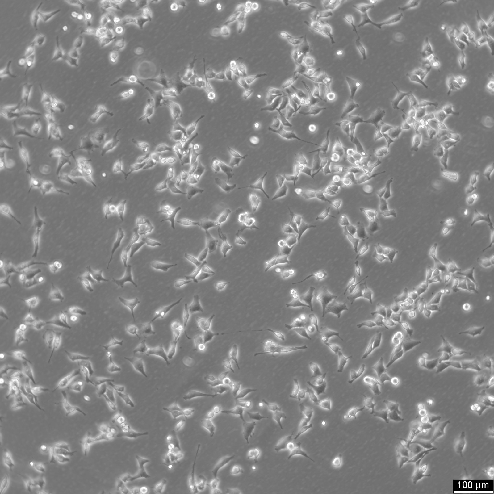

HK EGFP-alpha-tubulin/H2B-mCherry Cells
| Unit |
3 x 10^6 cells/ml |
|---|---|
| Cell Type |
Stable (KO/Expressing) Cells |
| Tissue Type |
Reporter-labeled cells |
| Organism |
Human |
| Donor History |
Carcinoma |
| Growth Properties |
Monolayer ,adherent |
| Growth Conditions |
Culture medium: DMEM ,w: 4.5 g/L Glucose ,w: 4 mM L-Glutamine ,w: 3.7 g/L NaHCO3 ,w: 1.0 mM Sodium pyruvate (Cytion article number 820300a). Supplements: Supplement the medium with 10% FBS. Dissociation reagent: Accutase. Seeding density: 1 x 10⁴ cells/cm². Freeze medium: As a cryopreservation medium ,we use complete growth medium (including FBS) + 10% DMSO for adequate post-thaw viability ,or CM-1 (Cytion catalog number 800100) ,which includes optimized osmoprotectants and metabolic stabilizers to enhance recovery and reduce cryo-induced stress. Incubation atmosphere: 37°C ,5% CO2 ,humidified atmosphere. |
| Immortalization Method | |
| Full Information |
https://www.cytion.com/HK-EGFP-alpha-tubulin-H2B-mCherry-Cells/300670 |

The HK EGFP-alpha-tubulin/H2B-mCherry HeLa Kyoto cell line is a meticulously engineered model designed for detailed visualization of cellular processes. This clonal line has been stably transfected to express two fluorescent protein fusions that enable real-time imaging of both chromatin and the microtubular network. The red fluorescent protein mCherry is fused to the core histone protein H2B, creating H2B-mCherry. This fusion protein is expressed from the pH2B-mCherry-IRES-neo3 plasmid and serves as a chromatin marker, highlighting the nuclear DNA in live-cell imaging and facilitating studies on chromatin dynamics and nuclear architecture.
Additionally, this cell line expresses monomeric enhanced GFP (Green Fluorescent Protein) fused to α-tubulin, introduced via the pmEGFP-α-tubulin-IRES-puro2b plasmid. The GFP-α-tubulin fusion provides a vivid green fluorescence that outlines the microtubule structures within the cell. This feature is crucial for studying microtubule organization, dynamics, and their role in cell division and intracellular transport. The stable integration of these constructs allows for continuous, long-term observation of these cellular components without the need for repeated transfection, thus reducing variability and enhancing the reliability of experimental results. Drug resistance selection following transfection ensures the stability and uniformity of expression among the cells in this line.
Společnost Cytion vznikla v roce 2004 v německém Heidelbergu s jasným vědeckým posláním: dodávat ověřené lidské a zvířecí buněčné linie bez kontaminace, připravené za přísně kontrolovaných laboratorních podmínek.
Postupně se vypracovala ve spolehlivou buněčnou banku, která slouží akademickým laboratořím, biotechnologickým firmám i farmaceutickým výzkumným týmům po celém světě. Klade přitom důraz na reprodukovatelnost výsledků, přísnou kontrolu kvality a dlouhodobá partnerství.