
 Genetika člověka
 Mikrobiologické aplikace
 NGS
 Rapid testy

 PCR plasty
 Pipetovací špičky
 Plasty pro buněčnou kultivaci
 Systém pro dlouhodobé skladování vzorků
 Zkumavky

 Třepačky
 Suché lázně
 Centrifugy
 End Point cyclery
 Real-time PCR cyclery
 Fluorometry
 Spektrofotometry
 Izolátory nukleových kyselin
 Pipetovací roboty
 Biobanking
 Live Cell Imaging

 Jednokanálové
 Multikanálové
 Sérologické pipety
 Jednorázové pipety

 Buněčné kultury
 PCR, qPCR, RT
 Detekce Mycoplasmat
 Reverzní transkripce

 Alergie
 Intolerance
 Elisa
 Imunologické analyzátory
 Veterinární imunologie
 Alergie u zvířat
 Intolerance u zvířat

 Čtečky identifikačních čipů
 Termočipy
 Biochemie
 Imunoanalýza


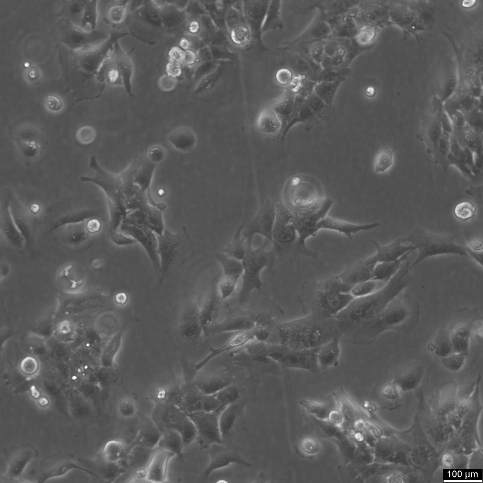

HMC3 Cells
| Unit |
3 x 10^6 cells/ml |
|---|---|
| Cell Type |
Immortalized Cells |
| Tissue Type |
Brain cancer cell lines |
| Organism |
Human |
| Donor History | |
| Growth Properties |
Adherent |
| Growth Conditions |
Culture medium: DMEM:Ham's F12 (1:1) ,w: 3.1 g/L Glucose ,w: 2.5 mM L-Glutamine ,w: 15 mM HEPES ,w: 0.5 mM Sodium pyruvate ,w: 1.2 g/L NaHCO3 (Cytion article number 820400a). Supplements: Supplement the medium with 10% FBS. Dissociation reagent: Accutase. Freeze medium: As a cryopreservation medium ,we use complete growth medium (including FBS) + 10% DMSO for adequate post-thaw viability ,or CM-1 (Cytion catalog number 800100) ,which includes optimized osmoprotectants and metabolic stabilizers to enhance recovery and reduce cryo-induced stress. Incubation atmosphere: 37°C ,5% CO2 ,humidified atmosphere. |
| Immortalization Method | |
| Full Information |

The Human Microglial Clone 3 (HMC3) cell line was developed in 1995 by Professor Tardieu’s team through the SV40-dependent immortalization of microglial cells from human spinal cord and cortical tissues, obtained from embryos aged between 8 to 12 weeks. These primary cells, characterized by slow division and complex morphologies, were initially cultured for 10-15 days before immortalization. The HMC3 cells maintained several key features of primary microglia, such as a diverse expression of myeloid markers like CD68, CD11b, and CD14, though the expression levels varied notably with the choice of primary antibody, particularly for CD68.
Following immortalization, the HMC3 cells exhibited enhanced proliferation rates, with doubling times between 24 and 48 hours, while preserving many phenotypic and morphological characteristics of their primary counterparts. Notably, there was a higher proportion of CD68 EBM/11-positive cells and a reduction in phagocytic activity compared to the primary cells. Stability in antigenic expression was confirmed across 35 passages, with the cells remaining positive for NSE, CD68, and CD11b, but negative for CD14, MHCII, and CD4 under baseline conditions. However, exposure to interferon-γ (IFNγ) elevated MHCII expression, aligning more closely with primary culture responses to the same treatment.
Functionally, the HMC3 line distinguished itself by producing higher levels of interleukin-6 (IL-6) under basal conditions compared to other clones. Despite this, a direct comparison with primary microglial cells’ cytokine production remains challenging due to methodological differences. The response to lipopolysaccharide (LPS) stimulation in these immortalized lines appeared diminished relative to primary cultures. Consistent with primary microglial characteristics, the HMC3 and other cloned lines did not produce tumor necrosis factor-alpha (TNFα), either spontaneously or following pro-inflammatory stimulation, highlighting a specific trait of human embryonic microglia.
Společnost Cytion vznikla v roce 2004 v německém Heidelbergu s jasným vědeckým posláním: dodávat ověřené lidské a zvířecí buněčné linie bez kontaminace, připravené za přísně kontrolovaných laboratorních podmínek.
Postupně se vypracovala ve spolehlivou buněčnou banku, která slouží akademickým laboratořím, biotechnologickým firmám i farmaceutickým výzkumným týmům po celém světě. Klade přitom důraz na reprodukovatelnost výsledků, přísnou kontrolu kvality a dlouhodobá partnerství.