
 Genetika člověka
 Mikrobiologické aplikace
 NGS
 Rapid testy

 PCR plasty
 Pipetovací špičky
 Plasty pro buněčnou kultivaci
 Systém pro dlouhodobé skladování vzorků
 Zkumavky

 Třepačky
 Suché lázně
 Centrifugy
 End Point cyclery
 Real-time PCR cyclery
 Fluorometry
 Spektrofotometry
 Izolátory nukleových kyselin
 Pipetovací roboty
 Biobanking
 Live Cell Imaging

 Jednokanálové
 Multikanálové
 Sérologické pipety
 Jednorázové pipety

 Buněčné kultury
 PCR, qPCR, RT
 Detekce Mycoplasmat
 Reverzní transkripce

 Alergie
 Intolerance
 Elisa
 Imunologické analyzátory
 Veterinární imunologie
 Alergie u zvířat
 Intolerance u zvířat

 Čtečky identifikačních čipů
 Termočipy
 Biochemie
 Imunoanalýza


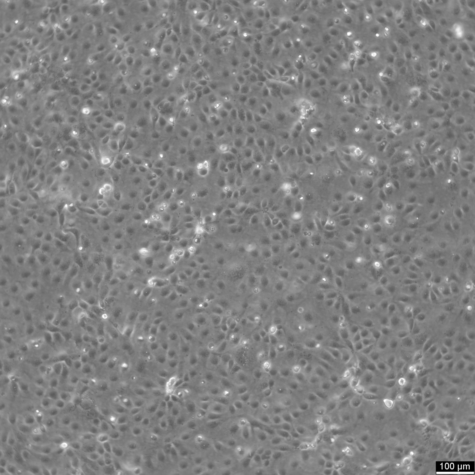

HCE-T Cells
| Unit |
3 x 10^6 cells/ml |
|---|---|
| Cell Type |
Immortalized Cells |
| Tissue Type |
Transformed cell lines |
| Organism |
Human |
| Donor History | |
| Growth Properties |
Adherent |
| Growth Conditions |
Culture medium: DMEM:Ham's F12 (1:1) ,w: 3.1 g/L Glucose ,w: 2.5 mM L-Glutamine ,w: 15 mM HEPES ,w: 0.5 mM Sodium pyruvate ,w: 1.2 g/L NaHCO3 (Cytion article number 820400a). Supplements: Supplement the medium with 5% FBS ,1% ITS (0.625 mg/mL human insulin ,0.625 mg/mL human transferrin ,0.625 microgram/mL sodium selenite ,0.535 mg/mL linoleic acid ,125 mg/mL BSA) and 10 ng/mL human EGF. Dissociation reagent: Accutase. Freeze medium: As a cryopreservation medium ,we use complete growth medium (including FBS) + 10% DMSO for adequate post-thaw viability ,or CM-1 (Cytion catalog number 800100) ,which includes optimized osmoprotectants and metabolic stabilizers to enhance recovery and reduce cryo-induced stress. Incubation atmosphere: 37°C ,5% CO2 ,humidified atmosphere. |
| Immortalization Method | |
| Full Information |

HCE-T is an SV40-transformed human corneal epithelial cell line derived from primary human corneal epithelium. The line was established by infection with a recombinant SV40–adenovirus hybrid vector (Ad–SV40), enabling stable expression of SV40 large T antigen and continuous proliferation. The original characterization specifically aimed to generate a continuously growing corneal epithelial cell line without shedding free viral particles.
In culture, HCE-T cells show typical epithelial “cobblestone” morphology and grow as adherent monolayers. Ultrastructural epithelial features such as desmosomes and apical microvilli have been reported, and the cells have been described as producing a cornea-associated 64 kD keratin. Under suitable differentiation conditions (e.g., air–liquid interface culture on collagen), HCE-T cells can form multilayered, stratified structures and develop measurable barrier properties, supporting their use in ocular surface research.
HCE-T cells are widely used to study corneal epithelial barrier function, permeability and formulation effects, migration/repair-related processes, and cellular responses to inflammatory or irritant stimuli. However, transporter expression patterns and differentiation-marker profiles can differ from native human cornea and from primary limbal/corneal epithelial systems. Therefore, HCE-T is best suited for mechanistic and comparative in vitro studies, while direct quantitative extrapolation to in vivo human corneal absorption or corneal differentiation biology should be performed with caution.
Společnost Cytion vznikla v roce 2004 v německém Heidelbergu s jasným vědeckým posláním: dodávat ověřené lidské a zvířecí buněčné linie bez kontaminace, připravené za přísně kontrolovaných laboratorních podmínek.
Postupně se vypracovala ve spolehlivou buněčnou banku, která slouží akademickým laboratořím, biotechnologickým firmám i farmaceutickým výzkumným týmům po celém světě. Klade přitom důraz na reprodukovatelnost výsledků, přísnou kontrolu kvality a dlouhodobá partnerství.